Value of Early Detection
TreVia’s solutions are making a significant impact in preventative care and early detection.
TreVia’s digital retinal imaging solutions provide early detection for a wide range of acute and systemic diseases. This level of preventative care ensures early treatment and helps to prevent blindness and other preventable complications.
Below are descriptions and images of the three most common conditions detected by TreVia technologies.
Diabetic Retinopathy
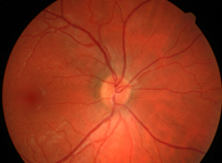 |
Normal Retina | 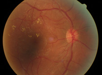 |
Diseased Retina |
| Normal Retina | Diseased Retina |
Diabetic retinopathy is the leading cause of preventable blindness among U.S. adults. The term “diabetic retinopathy” refers to all disorders of the retina caused by diabetes. There are two major types of retinopathy: nonproliferative and proliferative.
Nonproliferative retinopathy is a condition when the capillaries in the back of the eye balloon and form pouches. This condition move through three stages: mild, moderate and severe.
Proliferative retinopathy is when an individual’s retinopathy is so far progressed that the blood vessels in the eye become damaged and closed off. In response, new blood vessels start growing in the retina. These new vessels are weak and can leak blood, blocking vision and even causing scar tissue to grow.
The image on the right demonstrates yellowish hard exudates from damaged blood vessels. The round yellowish optic disk also demonstrates overlying small frond-like vessels associated with neovascularization or proliferative diabetic retinopathy.
Left undetected, diabetic retinopathy can lead to severe vision loss and blindness. The American Diabetes Association strongly recommends that all diabetics receive an annual eye exam along with an advanced screening to detect this condition.
Age related macular degeneration (AMD)
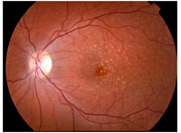 Age related macular degeneration (AMD) is a common disease often affecting those over 50 years that can lead to blindness. The macula is a sensitive portion of the retina that helps with central vision. AMD can be divided into early, intermediate and late forms. Another way to category AMD is dry or wet, where wet the form refers to neovascularization or proliferation of new blood vessels that can damage the adjacent retina. The wet or late form is often more severe and can progress more rapidly.
Early detection of AMD is critical because early AMD can be closely monitored for progression which allows intervention before irreversible damage occurs. Vitamin therapy for the eye has been clinically proven to slow progression of AMD in patients with intermediate forms. Late AMD results in advanced vision loss which can continue to progress even with treatments designed to decrease vessel proliferation such as injection therapy or laser surgery.
AMD screening can be performed with vision tests designed to check for central vision loss such as the Amsler grid. Digital retinal imaging can directly assess the retina and macular for protein deposits (called drusen) and vessel growth. Optical coherence tomography (OCT) uses ultrasound technology to assess damage to the retina.
The image above demonstrates small yellow white flecks of protein deposits called drusen around the macula.
Glaucoma
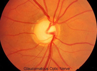 Glaucoma is a group of disorders that affect vision and can lead to blindness. Glaucoma damages the optic nerve in the back of the eye. It is usually related to abnormal circulation of the natural fluid (aqueous humour) within the eye. Processes which alter this normal flow of fluid can increase the pressure within the eye (intraocular pressure or IOP) which then damages the optic nerve. While most often related to increased intraocular pressure, there are forms of glaucoma in which the pressure is not elevated. Patients with glaucoma may be asymptomatic for some time before vision loss occurs. That is why screening is important to identify these patients so that intervention or treatment with medicine or surgery can be performed before irreversible damage occurs.
This image demonstrates a swollen, enlarged, and edematous optic disc associated with nerve damage from glaucoma.
Glaucoma can be detected with simple vision tests and pressure measurements called tonometry. Tonometry is simple and easy to perform.
